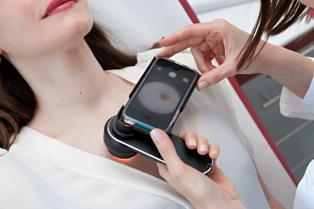
The global public health problem today becomes skin cancer, especially being one of the deadliest cancers: early detection is important, as early intervention improves survival. Skin cancer screening traditionally used to be dermatological examinations and biopsies, but recent advances in AI are changing that. AI-powered smartphone applications and imaging devices offer potential non-invasive, accessible, and cost-effective tools for assessing the risk of melanoma. The present review focuses on the role of AI in skin cancer screening, looking into the capabilities, limitations, and future prospects of AI-based diagnostic technologies.
The Growing Burden of Skin Cancer
Melanoma is a skin cancer with relatively low occurrence but proves to be the most fatal, taking the lion’s share of deaths due to skin cancer (Whiteman et al., 2016). The great risk is mostly UV radiation, so their early detection is the prime determinant in reducing deaths. Though the clinical examination of suspected melanoma is still being performed according to ABCDE parameters (Asymmetry, Border irregularity, Color variation, Diameter, and Evolution), technologies are being developed today to make this process more accurate (Esteva et al., 2017).
AI-Powered Smartphone Applications for Skin Cancer Screening
Some smartphone apps for the assessment of skin by AI have begun to emerge, which help people assess a suspected melanoma on their skin. These use deep learning algorithms trained on a large dataset of skin lesion image samples to analyze moles and give some risk assessment. Among AI apps are:
- SkinVision: AI algorithms analyze mole images and classify them as being at low, medium, or high risk for melanoma. Clinically validated, the app has an 80% sensitivity for skin cancer detection (Maier et al., 2020).
- MiiSkin: monitors moles over time for changes with an AI platform that detects irregularities.
- DermaScan: AI risk assessment of suspicious moles and recommendations regarding follow-up care.
These apps increase the opportunity for users to monitor their skin health on a regular basis, thus avoiding a lot of dermatology visits, enabling them to detect any suspicious lesions early.
AI-Driven Imaging Devices for Dermatology
The AI-powered imaging devices used in melanoma screening have improved accuracy beyond just smartphone apps. These devices are mostly used in dermatology clinics and combine high-resolution imaging with AI algorithms to detect the early signs of melanoma.
- DermoScan: AI-assisted dermoscopy analyzing skin lesions quantitatively with higher precision than the human eye.
- MoleScope: A miniaturized dermatoscope attached to the smartphones for well-moderated AI analysis, zooming, and capturing and detailing skin lesions in high definition.
- FotoFinder: The complete body-mapping means an AI algorithm-based assessment of moles over time.
This seamless integration creates a continuum between consumer-versatile applications and clinical-grade diagnostic instruments for the improvement and efficiency of melanoma screening.
How AI Algorithms Work in Skin Cancer Detection
Skin cancer screening AI models are mainly based on convolutional neural networks (CNNs), a specific variety of deep learning architecture aimed for image recognition. Such networks are trained on large volumes of datasets with thousands of images of benign and malignant skin lesions. The AI distinguishes normal moles from high-risk lesions based on visual clues that might not be obvious to the human eye and which were trained from an image from the thousands of examples the AI was fed into it (Esteva et al., 2017). The process for AI screening usually involves the following:
- Image Capture: The user photographs a mole with the mobile camera, an in-house imaging device, etc.
- Treatment: The AI holds the image from some irrelevant backgrounds and so on with different lighting conditions.
- Feature Extraction: The algorithm manages the characteristics of lesions according to shape, color, and texture.
- Risk Assessment: The AI matches the mole’s features against a database of known moles and then categorizes the risk level of the lesion.
- Recommendation: The system’s output is feedback on whether or not the user would benefit from a dermatological consultation.
Advantages of AI-Enhanced Skin Cancer Screening
Benefits of using AI-powered screening tools include:
- Access: This has allowed patients to check their skins from the comfort of their homes – breaking down barriers to early detection.
- Affordability: The AI diagnosis is cheap for frequent dermatologist visits and invasive tests.
- Timely diagnosis: Melanoma detection early by AI algorithms improves possible chances of good outcomes by the patient.
- Time-saving: This means faster diagnosis for dermatologists to get time for more high-risk patients.
Limitations and Challenges
While AI has proven effective for skin cancer diagnosis, some challenges include;
- Misclassifications: False negatives as well as false positives cause unnecessary worry for patients, whereas some cases go unnoticed.
- Non-standardized Applications: AI tools have no universally applicable standards when it comes to applications.
- Quality of the Record: An AI program cannot function if poor illumination, low-quality cameras, or inappropriate angles are present.
- Ethical Concerns: Privacy issues are important in respect to recorded personal skin images since they can be breached in security protocols or confidentiality may be violated.
Future Directions and Research
The AI evolution, meanwhile, presents constant dynamics towards increasing accuracy and reliability in skin cancer detection. Within the few foreseeable advancements would be:
- Integration with Telemedicine: Teledermatology services could be associated with AI-based screening for remote dermatological consultations.
- Multimodal AI Approaches: Integrating AI image analysis with genetic and biomarker data would increase the precision of diagnosis.
- Federated Learning: A decentralized AI training method that allows an algorithm to enhance using multiple data sources, while maintaining the privacy of all users. (Nguyen et al., 2021).
Skin cancer screening augmented by AI is changing the face of dermatology. Melanoma detection is now more available and, more importantly, efficient. Smartphone apps and imaging devices deployed with AI algorithms help the user in self-auditing their skin health while assisting dermatologists in diagnosing potentially high-risk lesions. Some challenges relating to false negatives, false positives, image quality, and data privacy remain. Nonetheless, continued improvements in AI technology promise everlasting long-term return for early detection and management of melanoma. Future integration of AI in clinical practice and under some regulatory framework can help to dramatically reduce skin cancer-related mortality rates.
References
- Esteva, A., Kuprel, B., Novoa, R. A., Ko, J., Swetter, S. M., Blau, H. M., & Thrun, S. (2017). Dermatologist-level classification of skin cancer with deep neural networks. Nature, 542(7639), 115-118. https://doi.org/10.1038/nature21056
- Maier, T., Kulichova, D., Schotten, K., Koller, S., Zelger, B., Richtig, E., & Hofmann-Wellenhof, R. (2020). Accuracy of a smartphone application using fractal image analysis of pigmented moles compared to clinical diagnosis and histological result. Journal of the European Academy of Dermatology and Venereology, 34(4), 857-863. https://doi.org/10.1111/jdv.15907
- Nguyen, D. C., Ding, M., Pathirana, P. N., & Seneviratne, A. (2021). Federated learning for AI-enabled healthcare applications: A taxonomy, current trends, and research challenges. Journal of Biomedical Informatics, 113, 103634. https://doi.org/10.1016/j.jbi.2020.103634
- Tschandl, P., Rinner, C., Apalla, Z., Argenziano, G., Codella, N., Halpern, A., … & Kittler, H. (2019). Human-computer collaboration for skin cancer recognition. Nature Medicine, 25(8), 1220-1224. https://doi.org/10.1038/s41591-019-0503-7
- Whiteman, D. C., Green, A. C., & Olsen, C. M. (2016). The growing burden of invasive melanoma: Projections of incidence rates and mortality to 2031 in Australia. Journal of Investigative Dermatology, 136(6), 1161-1169. https://doi.org/10.1016/j.jid.2016.01.035


